Fragmented Medial Coronoid Process (FMCP)
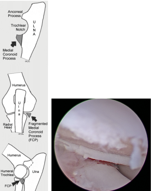
A fragmented medial coronoid process (FMCP) is a developmental disorder of the elbow that occurs when part of the medial coronoid process of the ulna becomes malformed, damaged, or separates from the surrounding bone.
The elbow is a complex joint made up of the humerus, radius, and ulna. When these bones do not fit together properly, abnormal forces are placed on the joint. This can damage the cartilage and lead to inflammation, pain, and progressive osteoarthritis.
FMCP commonly affects both elbows, though it is often worse on one side. Signs often begin between five and twelve months of age and may include forelimb lameness, elbow pain, stiffness, or worsening lameness after exercise or periods of rest. When both elbows are affected, lameness may be less obvious because the dog is compensating on both sides.
Diagnosing FMCP
Diagnosis begins with an orthopedic examination. Our surgeons evaluate your dog's gait and assess the elbows for swelling, pain, decreased range of motion, and other abnormalities. X-rays may be performed to see if there are changes associated with elbow dysplasia and osteoarthritis. A CT scan is the most effective method to evaluate the elbow because it allows our surgeons to identify the fragments and any abnormalities or changes with the bone.
Treatment of FMCP
When appropriate, early surgical treatment can help address the source of irritation within the joint and may help slow the progression of osteoarthritis.
Our surgeons typically perform an arthroscopic examination of the elbow, allowing direct visualization of the joint and cartilage. The fragmented or damaged portion of the medial coronoid process can then be removed, along with damaged cartilage and other abnormal tissue when indicated.
Following surgery, controlled activity and a structured rehabilitation plan are important parts of recovery. The long-term outlook depends largely on the amount of osteoarthritis present. Even when FMCP is diagnosed and treated early, some degree of arthritis may develop over time and may require ongoing management.
Osteochondritis Dissecans (OCD)
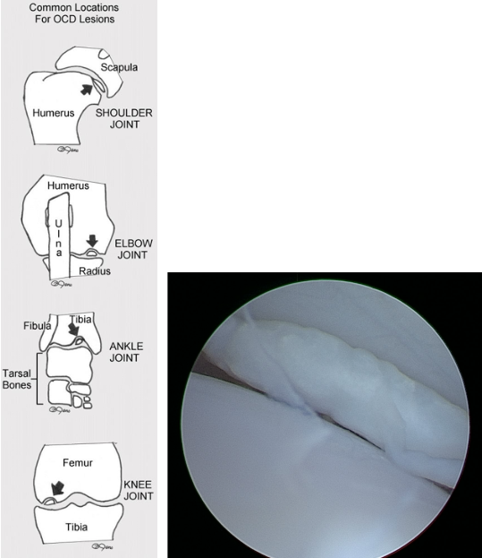
Osteochondritis dissecans (OCD) is a developmental disorder that affects the cartilage covering the surface of a joint and the underlying bone.
During normal growth, cartilage develops and is gradually replaced by bone. With OCD, an area of cartilage can become abnormally thick and does not receive adequate support from the underlying bone. Over time, cracks can develop within the cartilage, causing a portion to separate from the underlying bone and form a loose cartilage flap.
The damaged cartilage and exposed underlying bone can cause inflammation, pain, and lameness. Continued irritation can eventually lead to permanent osteoarthritis.
The shoulder is the most affected joint, although OCD can also occur in the elbow, stifle (knee), and hock (tarsus).
Diagnosing OCD
Diagnosis begins with a detailed medical history and comprehensive orthopedic examination. Dogs with OCD typically have lameness that can be localized to a specific joint, with pain often identified during flexion and extension of the affected joint. A CT scan can provide a more comprehensive evaluation of the joint and surrounding bone to help identify the cartilage flap.
Treatment of OCD
When surgery is recommended, our surgeons utilize arthroscopy to directly evaluate and treat the affected joint.
The loose or damaged cartilage flap is removed, and unhealthy tissue within the underlying bone is carefully removed until healthy bone is reached. This creates an environment that encourages new blood supply and the formation of reparative tissue within the defect.
Early diagnosis and treatment can help reduce ongoing joint irritation and may improve long-term comfort and function. However, the degree of osteoarthritis already present at the time of treatment can influence the long-term prognosis.