
A cranial cruciate ligament (CCL) tear is one of the most common causes of hind-limb lameness in dogs. The CCL is an important ligament within the knee that helps stabilize the joint during normal weight-bearing and activity. It functions similarly to the ACL in people.
When the CCL is torn, the knee becomes unstable. Instead of the bones moving normally together, the tibia (shin bone) can slide forward beneath the femur (thigh bone) when your dog puts weight on the leg. This abnormal movement causes pain and inflammation. It can also damage other structures within the knee, including the meniscus. Over time, continued instability can lead to progressive arthritis and chronic lameness.
CCL disease can affect dogs of any breed, size, or age. In many dogs, the ligament gradually weakens over time rather than tearing from one specific traumatic event.
Diagnosis
Diagnosing a CCL tear begins with a thorough history and orthopedic examination. Many dogs present with sudden or gradually worsening lameness in one rear leg. Some dogs may initially refuse to bear weight on the limb, while others develop a more subtle or intermittent limp.
During the orthopedic examination, we assess the knee for instability, swelling, pain, and changes in range of motion. A specific test called the cranial drawer test evaluates abnormal forward movement of the tibia relative to the femur. This movement is normally prevented by the CCL. In some patients, sedation or anesthesia may be necessary to fully evaluate the knee.
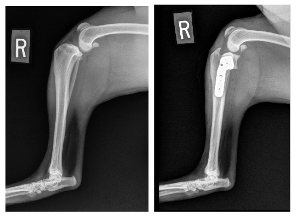
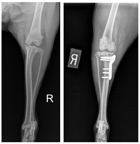
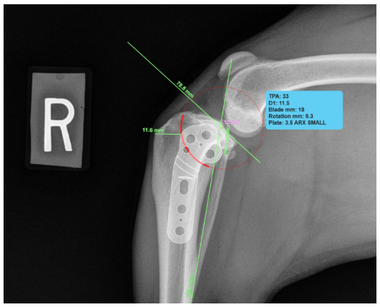
X-rays help us assess the knee for swelling, arthritis, and other bone or joint abnormalities and are also essential for planning TPLO surgery. When indicated, arthroscopy may be performed to directly evaluate the inside of the knee and identify damage to the meniscus, cartilage, and other joint structures.
Why Does a CCL Tear Need Treatment?
An unstable knee does not simply heal back to normal on its own. Although scar tissue can develop around the joint and provide some additional support, the underlying instability can continue to cause pain and contribute to arthritis.
For many dogs, surgical stabilization provides the best opportunity for a comfortable, functional knee.
TPLO Surgery
Tibial Plateau Leveling Osteotomy (TPLO) is an advanced surgical technique designed to stabilize the knee without replacing the torn CCL.
Rather than attempting to recreate the damaged ligament, a TPLO changes the angle of the top of the tibia so that the forces acting on the knee during weight-bearing are altered. This allows the knee to remain dynamically stable during normal activity despite the absence of a functioning CCL.
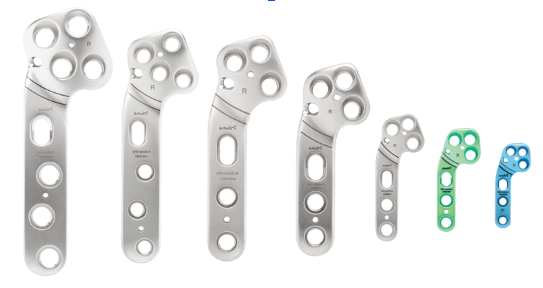
Before surgery, X-rays are taken and used to carefully measure the slope of the tibial plateau. During the procedure, our surgeon makes a precise curved cut in the upper portion of the tibia and rotates the tibial plateau to a predetermined angle. The bone is then secured in its new position with a specialized TPLO plate and screws.
As the bone heals, the corrected anatomy provides a stable, functional knee. The plate is typically designed to remain in place permanently.
Why TPLO Is the Gold Standard
Several surgical techniques exist for cranial cruciate disease, but they are not equal—and the differences matter for your dog. TPLO is considered by board-certified surgeons to be the gold standard for most dogs, and the objective, peer-reviewed evidence—measuring how dogs walk, how quickly they recover, how often complications occur, and how much arthritis develops—consistently backs that up. Here is why our team recommends it with confidence.
It Is a Fundamentally More Stable Repair
What truly sets TPLO apart is that it makes the knee stable through the dog’s own healed anatomy rather than relying on an implant or suture to stand in for the torn ligament. Once the bone heals, that stability is permanent, it does not depend on scar tissue forming or on flawless activity restriction during recovery, two factors that routinely undermine other techniques. This is a major reason TPLO performs so dependably in active dogs and in large and giant breeds.
It Delivers the Best-Documented Recovery
The results are hard to argue with. Studies using objective force-plate analysis—which precisely measures how much weight a dog places on each leg—consistently show TPLO restoring limb function better than the alternatives, with reported return of limb function around 93 percent in one year. In several comparative studies, TPLO was the only technique in which dogs reliably returned to normal function at a trot. In practical terms, most dogs go back to running, hiking, and playing.
It Is One of the Safest Options
TPLO is also one of the safest procedures available for this problem. A large systematic review found that, compared with tibial tuberosity advancement (TTA), TPLO had roughly half the rate of major complications and dramatically fewer catastrophic complications, along with fewer cases of ongoing joint instability. And because we inspect the joint thoroughly at the time of surgery—ideally with arthroscopy—any meniscal tear can be found and treated in the same procedure, which is one of the best ways to prevent a persistent limp down the road.
It Better Protects Against Future Arthritis
Because TPLO restores such stable, natural mechanics, it also helps slow the arthritis that inevitably follows cruciate disease. Systematic reviews of veterinary literature report a lower increase in osteoarthritis after TPLO than after tibial tuberosity advancement. Less arthritis over the years means more comfort, better mobility, and a better quality of life for the rest of your dog’s life.
Why TTA and MMP Are Not Our First Choice
Tibial tuberosity advancement (TTA) and its variations, including the Modified Maquet Procedure (MMP), address the same forces as TPLO but through a different, less forgiving mechanism. In head-to-head analysis of the veterinary literature, these procedures carry substantially higher rates of major and catastrophic complications than TPLO, along with higher reoperation rates and more late meniscal tears. The MMP depends on a thin cortical “hinge” of bone that can crack or fracture, and some published series report major complication rates approaching one in three. These techniques have a place in experienced hands for selected patients, but the objective, published data do not show them matching—let alone beating—TPLO.
Why the Lateral Suture Falls Short
The lateral suture (extracapsular repair) is an older technique that places a suture outside the joint to act as a temporary artificial ligament while scar tissue forms. It can give acceptable results in small, quiet dogs, but it has significant limitations: it depends entirely on that suture and on strict activity restriction holding up during healing, and in larger or active dogs the suture is subject to far greater force, so long-term durability is markedly lower than with TPLO. In a randomized, blinded clinical trial of 80 dogs, TPLO produced significantly higher weight-bearing and greater owner satisfaction one year after surgery, and more recent matched comparisons found TPLO functionally superior at every single time point up to a year. For active dogs and large breeds, the lateral suture is simply not a comparable option.
The Bottom Line for Your Dog
Every dog is different, and our surgeons always consider your dog’s size, age, activity level, tibial plateau angle, and any other joint problems before making a recommendation. In a small, quiet dog, a simpler repair can occasionally be appropriate. But for the great majority of dogs—and for essentially every active dog and large or giant breed—the evidence points clearly to TPLO as the procedure most likely to return your pet to comfortable, athletic function for life. When we recommend TPLO for your dog, it is because we believe it gives them the best possible outcome, and we are always glad to explain why.
What Can You Expect After TPLO?
A TPLO is designed to restore stability to the knee, reduce pain, and allow your dog to return to a normal, comfortable, and active lifestyle. As with any orthopedic surgery, successful recovery depends on careful postoperative management and allowing the bone adequate time to heal.
Your dog's activity will gradually increase throughout recovery at the surgeon’s direction, with follow-up examinations and X-rays used to monitor healing. Rehabilitation exercises are recommended to help restore muscle strength, joint motion, and normal use of the limb.
Because cranial cruciate disease is most often a gradual, degenerative process rather than a single injury, many dogs eventually develop a tear in the opposite knee as well. We will examine both hind limbs and talk with you about what to watch for, so a second problem can be caught and addressed early.
Our goal is not simply to repair an injured knee—it is to restore comfortable function and give your pet the best opportunity for an active, healthy life.
TPLO is the procedure most recommended by board-certified surgeons for cranial cruciate disease, particularly in larger and more active dogs. Most dogs regain excellent, comfortable limb function, and studies show that weight-bearing often returns to near-normal levels over the months following surgery. As with any orthopedic procedure, a small percentage of dogs experience complications such as incisional irritation, infection, or a later meniscal injury; these are usually manageable, and we work together with you to monitor them throughout recovery.